Figure 2.
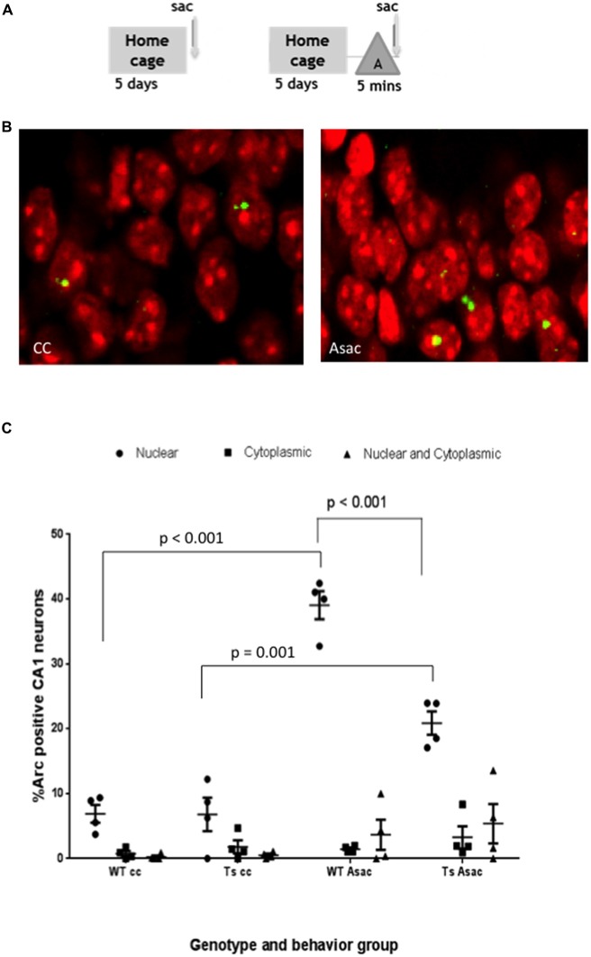
Pyramidal neurons in the CA1 hippocampal region of Ts65Dn mice express Arc mRNA in response to exploration of a novel environment but the percent of neurons expressing Arc is reduced compared to wild-type (WT) CA1. (A) Schematic of behavioral paradigm. Mice were taken from their home cage and immediately sacrificed (cage control, cc), or allowed to explore a novel enriched environment “A” for 5 min and then immediately sacrificed (Asac). (B) Representative confocal images from each behavioral condition showing transcription foci of Arc mRNA (yellow) within the nucleus (red). (C) Scatter plot showing the mean percent ± SEM of Arc mRNA positive neurons in area CA1 of WT and Ts65Dn (Ts) mice. The percent of neurons with intranuclear foci of Arc mRNA is greater in the Asac group compared to cc for both WT and Ts65Dn mice (WT p < 0.001, Ts p = 0.001). The percentage of neurons with intranuclear foci of Arc mRNA in the Ts65Dn group is reduced compared to WT mice, p < 0.001. Four mice per genotype per group were used. Neurons counted per group were WT cc – 900, Ts cc – 340, WT Asac – 710, Ts Asac – 767.

