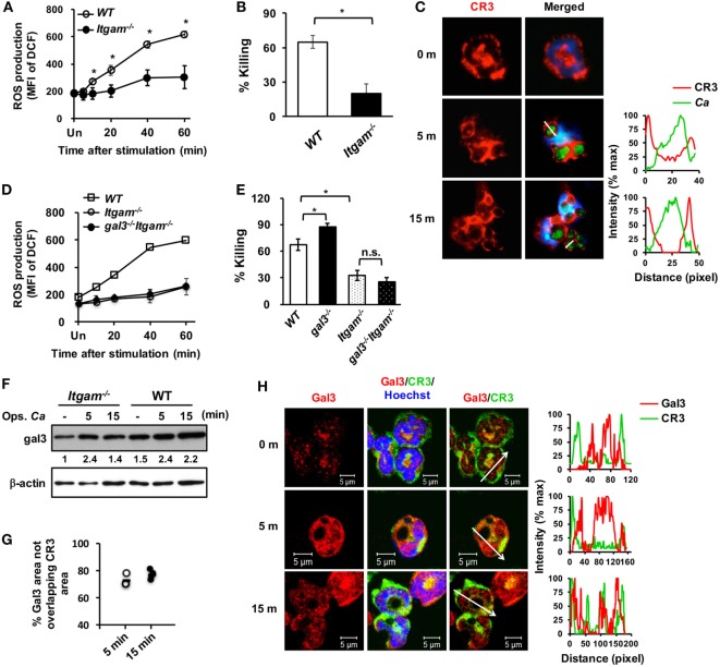
Figure 3.
Gal3 negatively regulates complement receptor 3 (CR3)-mediated killing of Candida by neutrophils. Neutrophils isolated from WT, gal3−/−, Itgam−/−, gal3−/−Itgam−/− mice were allowed to ingest opsonized Candida yeasts. (A,D) The level of reactive oxygen species (ROS) production is shown as mean fluorescence intensity (MFI) of oxidized DCF fluorescence. n = 3 in panel (A); n = 4 in panel (D). Experiment was performed twice. (B,E) The ability of neutrophils to kill opsonized Candida is shown as % killing. n = 3 in panel (B); and n = 4 in panel (E). Experiment was performed twice. Data are presented as mean ± SD. *p < 0.05; n.s., not significant, as analyzed by Mann–Whitney test. (C) Confocal fluorescence microscopic analysis to localize CR3 (red) and Candida OG1 (green). (F) Gal3 expression in Itgam−/− and WT neutrophils stimulated with opsonized Candida (Ops. Ca) was detected by Western blot. β-actin was used as a loading control. The numbers below the gel denote the relative intensity of gal3. Experiment was performed three times. (G,H) Confocal microscopy analysis to localize gal3 (red) and CR3 (green). The intensity of different fluorochromes along the white arrow in the merged (C) and Gal3/CR3 (H) images is shown in the histogram on the right. (G) Metamorph software was employed to determine the percentages of non-localization between gal3 and CR3 in the whole gal3 area in image in panel (H) (56,444 µm2 in average). Each dot represents one photo. n = 4.

