Figure 1.
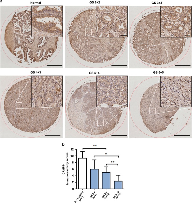
CRMP1 is downregulated in prostate cancer tissues. (a) CRMP1 immunohistochemistry. Representative micrographs show the CRMP1-immunostained tissue microarray spots of normal and malignant prostatic tissues. Magnification, × 40; bars, 500 μm. Insets show the indicated areas at higher magnification. Magnification, × 400; bars, 50 μm. The normal prostatic epithelial cells exhibited moderate granular immunostaining of CRMP1 in their cytoplasm. The malignant cells in low and moderately differentiated adenocarcinoma lesions (Gleason scores 2–7) showed weak cytoplasmic immunoreactivity, and showed barely detected or negative immunoreactivity in high-grade poorly differentiated lesions (Gleason scores 8–10). (b) CRMP1-IRS analysis performed on malignant and non-malignant (normal or benign prostatic hyperplasia) prostatic tissues. IRS scores were acquired by multiplying the level of staining intensity (negative=0, weak=1, moderate=2, strong=3) and percentage of positively stained cells (0%=0, <10%=1, 11–50%=2, 51–80%=3, >80%=4). Results showed that adenocarcinoma lesions of higher Gleason scores (Gleason score⩾5) showed significantly lower CRMP1 expression than normal or BPH tissues. *P<0.05; **P<0.01. IRS, immunoreactivity score.

