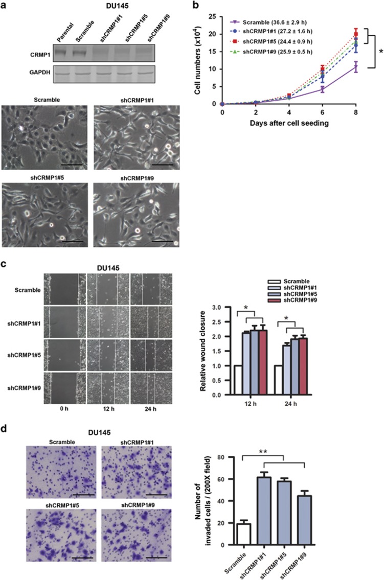
Figure 2.
CRMP1 knockdown promotes cell proliferation, migration and invasion capacities of prostate cancer cells. (a) Top: Representative immunoblots validated that DU145-shCRMP1 infectants showed very weak or barely detectable CRMP1 expression, whereas the parental and DU145-shScramble infectants expressed a moderate level of endogenous CRMP1. Bottom: Representative phase-contrast images of DU145-shCRMP1 and DU145-shScramble infectants. DU145-shCRMP1 infectants appeared more spindle-shaped and less adhered to one another at subconfluence. Magnification, × 20; bars, 1 μm. (b) Proliferation assay by cell counting. DU145-shCRMP1 infectants proliferated at faster rates than the shScramble infectants. Brackets show the doubling time (hours). (c) Wound healing assay. Left: Representative images of scratched and recovering of wounded areas (marked by solid lines) on confluence monolayers of shCRMP1- and shScramble infectants taken at different time points. Right: Semiquantitative analysis of wound closure. Relative wound closure was determined by measuring the width of the wounds. Data are presented as fold changes relative to the width of the wounds made by control shScramble infectants. DU145-shCRMP1 infectants showed significantly higher migration capacity than the DU145-shScramble infectants. (d) Matrigel invasion assay. Left: representative micrographs of the crystal violet-stained migrated shCRMP1- and shScramble infectants. DU145-shCRMP1 infectants showed significantly higher invasion capacity than the DU145-shScramble infectants. Magnification, × 20; bars, 1 μm. *P<0.05; **P<0.01.

