Figure 5.
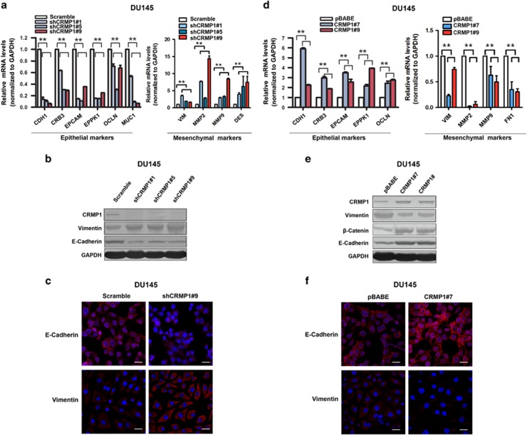
CRMP1 knockdown induces EMT but its overexpression induces mesenchymal–epithelial transition in DU145 prostate cancer cells. Real-time PCR, immunoblot and immunofluorescence analyses of EMT markers in (a–c) DU145-shCRMP1 and (d–f) DU145-CRMP1 infectants. (a, d) Real-time PCR analysis. Results showed that (a) DU145-shCRMP1 infectants expressed significantly lower levels of epithelial cell markers, including CDH1, CRB3, EPCAM, EPPK1, OCLN and MUC1, but higher levels of mesenchymal cell markers, including VIM, MMP2, MMP9 and DES, whereas (d) DU145-CRMP1 infectants expressed higher levels of epithelial cell markers but lower levels of mesenchymal cell markers (VIM, MMP2, MMP9 and FN1). Expression levels were normalized to glyceraldehyde 3-phosphate dehydrogenase (GAPDH). Data were presented as the mean±s.d. obtained from three independent experiments. **P<0.01 DU145-shCRMP1 or DU145-CRMP1 infectants versus shScramble- or pBABE infectants. (b, e) Immunoblot analysis. A significant reduced E-cadherin expression but an increased vimentin expression was shown in (b) DU145-shCRMP1 infectants as compared with shScramble infectants, whereas a decreased vimentin expression but increased expressions of E-cadherin and β-catenin were revealed in (e) DU145-CRMP1 infectants as compared with pBABE infectants. (c, f) Immunofluorescence. A significant reduction of E-cadherin but increase of vimentin immunosignals were detected in (c) DU145-shCRMP1 infectants, whereas an increase of E-cadherin but a reduction of vimentin immunosignals were seen in (f) DU145-CRMP1 infectants. Nuclei were counterstained by 4′,6-diamidino-2-phenylindole, dihydrochloride (DAPI). Magnification, × 400; bars, 50 μm.

