Figure 7.
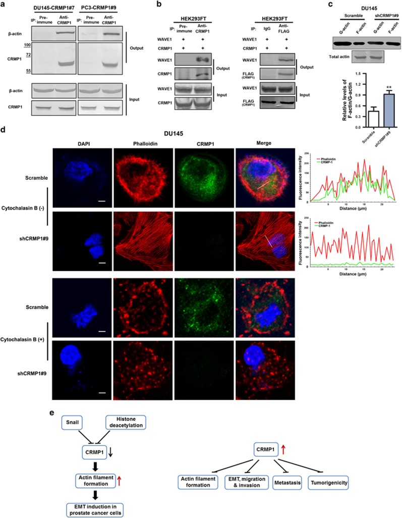
CRMP1 is associated with actin and WAVE1 and its knockdown promotes cytoskeletal actin filament formation in prostate cancer cells. (a) Immunoprecipitation (IP) performed in CRMP1 infectants of DU145 and PC-3 cells. Results showed that CRMP1 antibody but not pre-immune rabbit serum could immunoprecipitate β-actin in both DU145-CRMP1 and PC-3-CRMP1 infectants. (b) IP performed in HEK293 cells co-transfected with CRMP1 and WAVE1. Results showed that both anti-CRMP (left) and anti-FLAG (right) antibodies could immunoprecipitate WAVE1 in transfected HEK293FT cells. (c) F-actin/G-actin in vivo assay. Upper: representative immunblots show the monomeric G-actin and polymeric F-actin expressed in DU145-shCRMP1 and DU145-Scramble infectants. The immunosignal intensities of F-actin relative to that of G-action were quantified (n=3 for shCRMP1-/Scramble infectants). Immunoblots of total actin from DU145-shCRMP1 and DU145-Scramble infectants used in F-actin/G-actin assay are shown. Lower: semiquantitative analysis of F-actin/G-actin ratio in DU145 infectants. Results showed that CRMP1 knockdown could enhance the F-actin/G-actin ratio in DU145-shCRMP1 infectants as compared with DU145-Scramble infectants. **P<0.01 versus Scramble infectants. (d) Combined rhodamine-phalloidin staining for F-actin and CRMP1 immunofluorescence in DU145-shCRMP1 and DU145-Scramble infectants with or without cytochalasin B treatment. Upper two panels: In the absence of cytochalasin B, phalloidin-stained subcortical actin bundles, which outlined the cell boundaries, were visualized in DU145-Scramble infectants. CRMP1 and phalloidin-stained F-actin shared partial co-localization to the cytoplasm of the DU145-Scramble infectants. Prominent phalloidin-stained F-actin filaments or stress fibers were clearly visualized in DU145-shCRMP1 but not DU145-Scramble infectants. Fluorescent images were analyzed for intensity profiles using software FV10-ASW (Olympus, Tokyo, Japan). Pearson's correlation coefficient was measured for co-localization correlation of intensity distributions between two channels, with 10 cells totally analyzed in each experiment. The white lines in the merged images indicate the portions of cells where fluorescence intensities were measured. Pearson's coefficient: DU145-shCRMP1, 0.018±0.005; DU145-Scramble, 0.486±0.135. Lower two panels: In the presence of cytochalasin B, phalloidin-stained F-actin filaments were not seen in DU145-shCRMP1 infectants. Magnification, × 600; bars, 10 μm. (e) Schematic diagrams illustrating the transcriptional repression of CRMP1 by Snail and histone deacetylation and also its inhibitory effects in malignant and metastatic growth of prostate cancer cells via its regulation of cytoskeletal actin organization and suppression of EMT.

