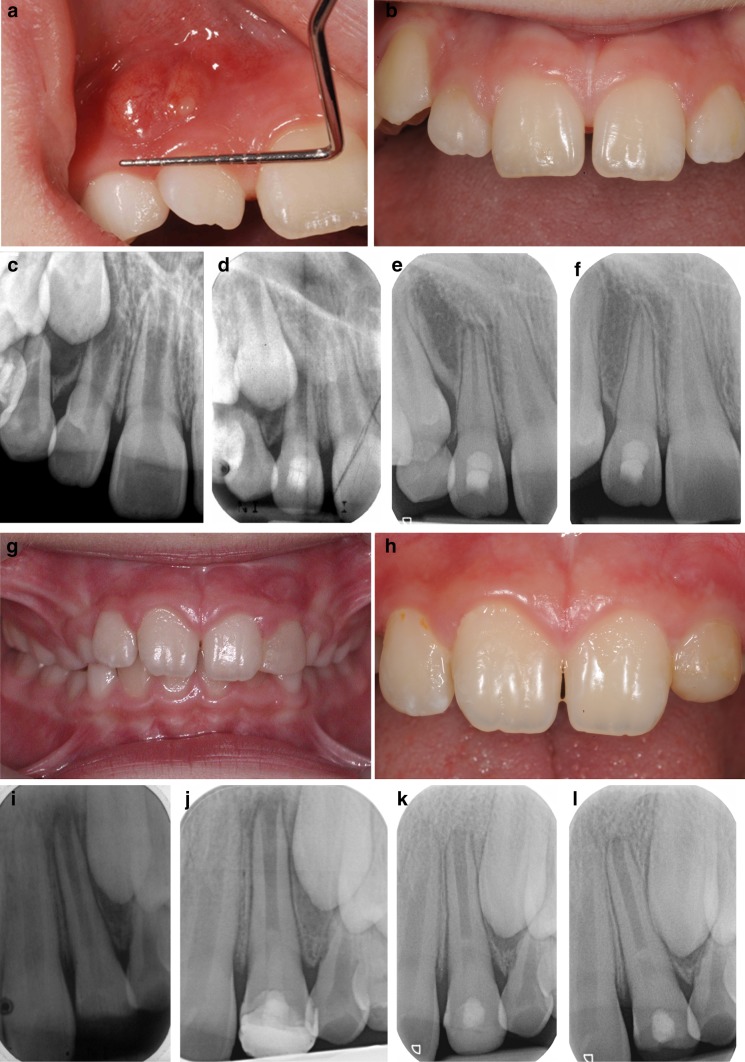
Fig. 2.
Clinical photographs and radiographic examination of two cases treated with regenerative endodontic technique showing success and survival outcomes. a Photograph showing labial abscess with discharging sinus related to the non-vital 12 secondary to dens invaginatus. b Photograph showing resolution of signs of infection (swelling and discharging sinus) maintained for up to 24 months following RET treatment of 12. c–f Periapical radiographs taken at baseline (showing an immature 12 with <1/2 root formation, thin dentinal walls and wide open apex), and follow-up at 3 months, 9 months and 2 years showing complete success following RET with gradual root formation and thickening of dentinal root walls. g Photograph showing traumatised non-vital 21 which sustained an enamel/dentine fracture. h Photograph showing no signs of infection (swelling and/or discharging sinus) at 24 months following RET treatment of 21. i–l Periapical radiographs taken at baseline (showing an immature 21 with <2/3 root formation, thin dentinal walls and wide open apex), and follow-up at 3 months, 9 months and 2 years showing no evidence of periapical lesion and with no signs of continuation of root development nor thickening of dentinal walls. The apical root canal space (l) shows evidence of radiopaque trabeculation suggestive of bony ingrowth

