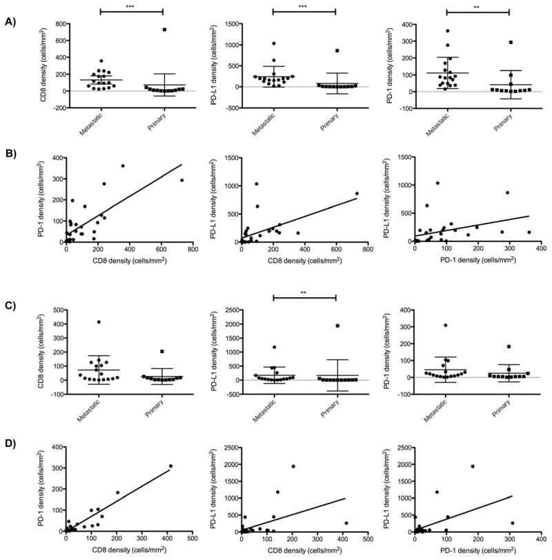
Figure 3.
A, Relative CD8, PD-L1, and PD-1 cell densities in the invasive margins of all samples of synovial sarcoma. PD-L1, PD-1, and CD8 cell densities were all significantly higher in the invasive margins of metastatic compared to primary tumors (**P <0.01, *** P <0.0001). Error bars represent standard deviations. B, Spearman correlation plots demonstrating that CD8, PD-L1, and PD-1 cell densities all positively correlate with one another within both the invasive margin C. Relative CD8, PD-L1, and PD-1 cell densities in the intra-tumor margins of all samples of synovial sarcoma. PD-L1 cell densities were significantly higher in the intra-tumor niche of metastatic compared to primary tumors (**P <0.01). D. Spearman correlation plots demonstrating that CD8, PD-L1, and PD-1 cell densities all positively correlate with one another within the intra-tumor niches.

