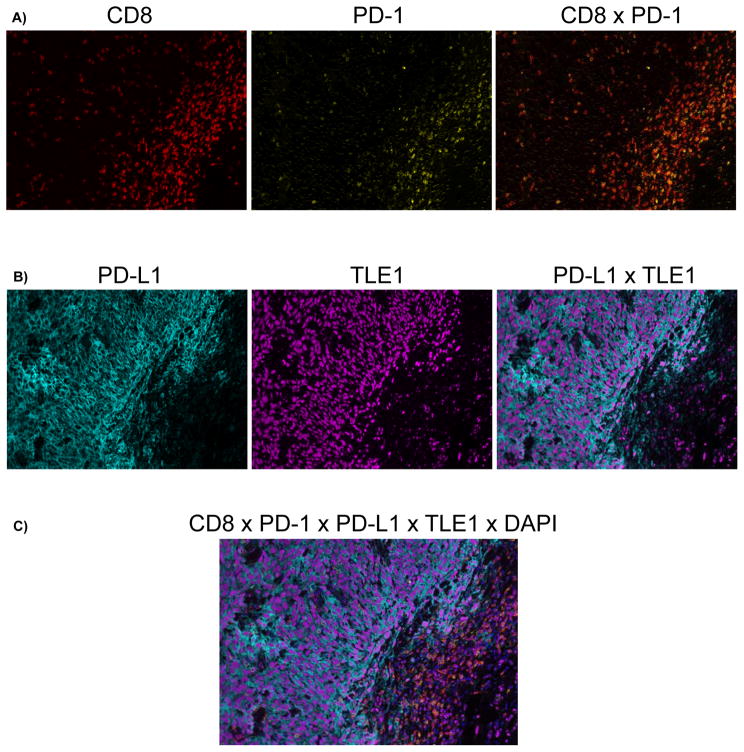
Figure 4.
Representative multiplex immunofluorescence image of synovial sarcoma with increased PD-L1/PD-1/CD8 cell densities at the tumor invasive margin demonstrating co-localization of PD-1/CD8 double-positive lymphocytes with PD-L1/TLE1 double-positive synovial sarcoma cells at the invasive margin. Color scheme - Red: CD8, Yellow: PD-1, Cyan: PD-L1, Magenta: TLE1, Blue: DAPI. A, Co-expression of CD8 and PD-1 on lymphocytes (Double positive = orange) concentrated at the invasive margin. B, Expression of PD-L1 by TLE1-positive synovial sarcoma cells at the invasive margin. C, Merged image of all labels. Magnification 200X.

