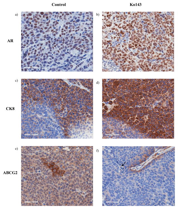
Figure 6. IHC analyses of the paraffin embedded tumor sections.
Tumors from control (n=8) and Ko143 (n=9) treated mice were dissected upon autopsy and a part of the tumors were subjected to paraffin embedding. The paraffin embedded sections were immunostained with (ab) AR, (c–d) CK8 and (e–f) ABCG2 and imaged under 40X magnification. Analysis was done in one section of the tumor from each animal.

