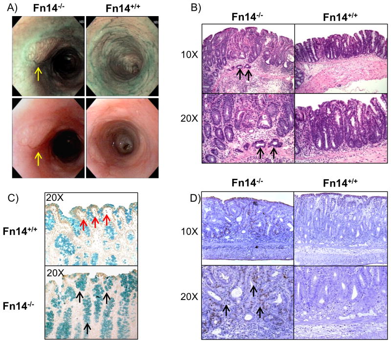
Figure 3. Fn14 protects from carcinogenesis induced by DSS-colitis.
(A) High-resolution endoscopic images of the distal colon during the recovery phase of DSS colitis (day 21) show tumor lesions in Fn14-/- mice, but no masses in Fn14+/+ mice (tumor lesions indicated by yellow arrows). (B) Representative histopathological sections show that cancer is present in Fn14-/- mice at the submucosal level (cancer cells indicated by black arrows). (C) TUNEL assay for apoptotic cells in colon sections of mice show that Fn14+/+ mice have an increased number of TUNEL-positive cells in comparison with Fn14-/- (apoptotic cells indicated by red arrows). The methyl green counterstaining shows that in the Fn14-/- mice there is an increased number of mucus secreting goblet cells (indicated by black arrows) in comparison with the Fn14+/+. (D) Mice were injected with BrdU 2 hr before sacrifice. Histological sections treated with BrdU antibody show that a higher number of cells were actively replicating their DNA at the level of the lamina propria (black arrows). Results are representative of two independent experiments.

