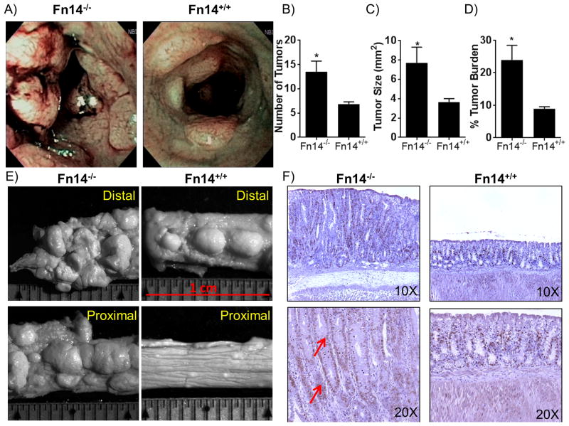
Figure 4. Susceptibility to carcinogenesis is enhanced in Fn14-/- mice in the AOM-DSS inflammation-driven colon cancer model.
(A) High-resolution endoscopic images of the distal colon after 1 injection of AOM followed by 2 cycles of 7 days of DSS plus 14 days of recovery show enhanced susceptibility of Fn14-/- mice (left panel) to carcinogenesis compared to Fn14+/+ mice (right panel), which showed fewer masses. (B) Quantification of the number of tumors showed that Fn14-/- mice develop a significant higher number of tumor compared to Fn14+/+ mice (unpaired t test; P < 0.05; n = 6/group). (C) The assessment of the size of the tumors shows that Fn14-/- mice developed tumors that were significantly bigger than Fn14+/+ mice (unpaired t test; P < 0.05; n = 6/group). (D) Tumor area expressed as a percentage of the whole colon shows that Fn14-/- mice develop a significantly greater tumor burden in comparison to Fn14+/+ mice (unpaired t test; P < 0.05; n = 6/group). (E) Images from stereomicroscopy examination of distal and proximal parts of colon of Fn14-/- and Fn14+/+ mice show a higher number of tumors in the Fn14-/- mice compared to the Fn14+/+ mice. (F) Histological sections treated with BrdU antibody show that a higher number of cells are actively replicating their DNA at the level of the lamina propria in Fn14-/- mice (red arrows) compared to Fn14+/+ mice. * P < 0.05, ** P < 0.01, *** P < 0.001. Results are representative of two independent experiments.

