
Figure 6. Intravenous administration of sTRAIL LPD inhibited the orthotopic desmoplastic BXPC3 tumor growth and remodeled the tumor microenvironments.
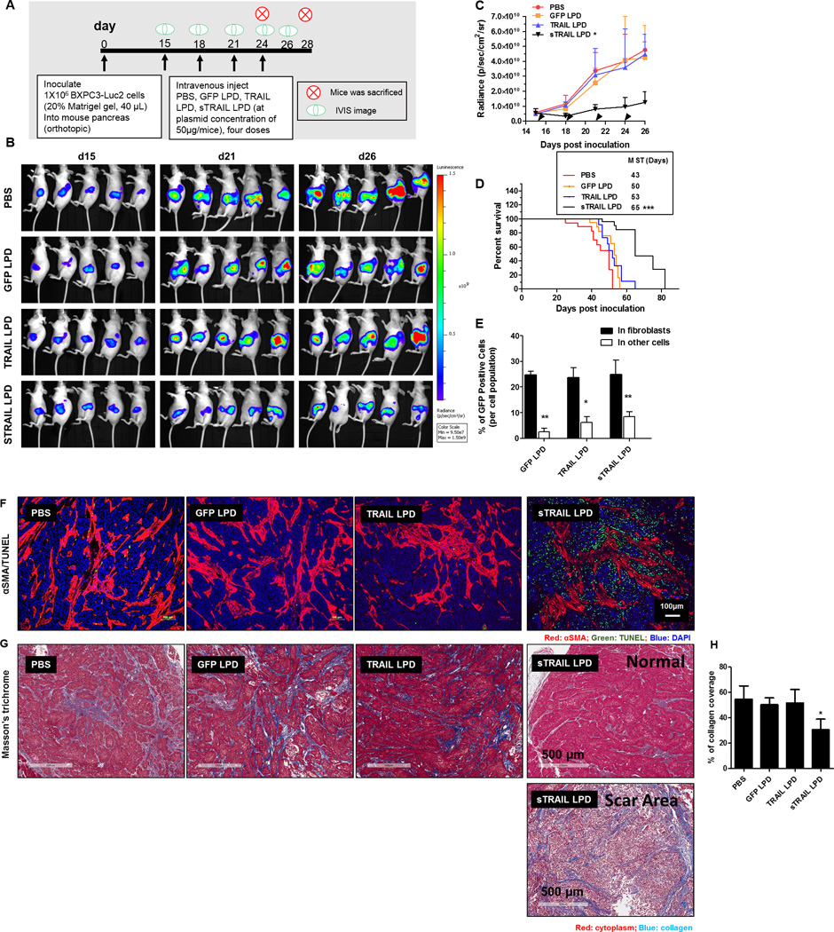
A. Dosing schedule of sTRAIL treatment on BXPC3-Luc2. B. IVIS images of BXPC3-Luc2 tumor after different treatments (n = 5). C. Tumor inhibition curve of BXPC3 (n = 6–10, *P < 0.05 compared to PBS group). (D) The survival proportions of the treated groups. Median survival time (MST) are presented in the inserted form (Data shown as mean ± SD, n= 6–8. ***, P < 0.001). E. Flow cytometry analysis of GFP’s association with αSMA positive fibroblasts 2 days after the third injection of the LPD (n = 4, * P < 0.05, ** P < 0.01). F. IF staining of αSMA and TUNEL assay from BXPC3-Luc2 tumor tissues after different treatments. G. Masson’s trichrome staining for collagen from the BXPC3-Luc2 tumors after different treatments. Heterogeneities are observed in the sTRAIL LPD groups. Scar tissue (with few cell structures) is observed. H. The quantification of collagen levels based on non-scar area (n = 4~5, * P < 0.05).