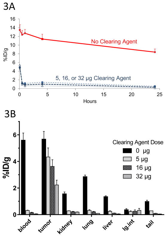
Figure 3. Optimization of DOTAY-Dextran CA Dose.
Figure 3A. Blood clearance of circulating C825-Fc-2H7 FP by various doses of DOTAY-Dextran CA. Mice were injected with 1.4nmol of the 2H7-Fc-C825 fusion protein FP followed by 0, 5, 16 or 32μg of DOTAY-Dextran CA 23-hours later. One hour later, 2.4nmol of 90Y-DOTA-Biotin was injected. Blood was collected after 0.08, 0.5, 1, 4, and 24-hours following injection of radioactivity. Results represent the calculated percentages of the injected dose per gram of tissue (%ID/g, ±SD; n=3) after corrections for decay and background subtraction (red square, diluent; filled circle, 5μg DYD; diamond, 16μg DYD; triangle, 32μg DYD).
Figure 3B. Biodistribution of 90Y-DOTA-Biotin in organs of mice bearing subcutaneous Ramos xenografts following injection of various doses of DOTAY-Dextran CA. Mice were injected with 1.4nmol of 2H7-Fc-C825 followed by 0, 5, 16, or 32μg of DOTAY-Dextran CA 23-hours later. One hour after injection of DOTAY-Dextran, 2.4nmol of 90Y-DOTA-Biotin was injected. Tissues were harvested 24-hours after the injection of radioactivity. Results represent the calculated mean percentages of the injected dose per gram of tissue (%ID/g, ±SEM; n=3) after corrections for decay and background.

