Figure 4.
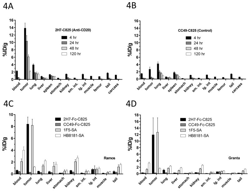
Figure 4A,B. Biodistribution of 90Y-DOTA-Biotin in mice bearing subcutaneous Ramos xenografts. Mice were injected with 1.4nmol of 2H7-Fc-C825 (A) or 1.4nmol of the control, non-binding CC49-Fc-C825 FP (B) followed by 5μg DOTAY-Dextran 23-hours later. One hour after the DOTAY-Dextran, 2.4nmol of 90Y-DOTA-Biotin was injected. Tissues were harvested 4, 24, 48 and 120-hours after injection of radioactivity. Results represent the %ID/g (±SEM; n=5) after corrections for decay and background subtraction.
Figure 4C, D. Biodistribution of 90Y-DOTA-Biotin in mice bearing subcutaneous Ramos xenografts (C) or Granta-519 xenografts. Mice were injected with 1.4nmol of 2H7-Fc-C825 (anti-CD20 bispecific), CC49-Fc-C825 (non-binding control bispecific antibody), 1F5-SA (anti-CD20-streptaviding conjugate) or HB8181-SA (non-binding control antibody-streptavidin conjugate), followed 23-hours later by either 5μg DOTAY-Dextran CA (for bispecific antibodies) or 5.8nmol NAGB CA (for antibody-streptavidin conjugates). One hour after injection of the CA, 2.4nmol of 90Y-DOTA-Biotin was administered. Tissues were harvested 4, 24, 48 and 120-hours after injection of radioactivity. Results represent the calculated %ID/g(±SEM; n=5) after corrections for decay and background subtraction.

