Figure 6.
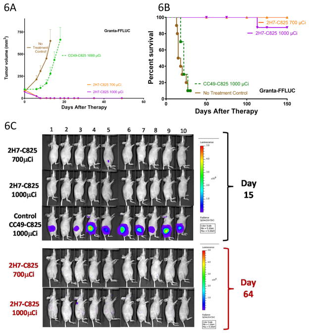
Figure 6A, B. Tumor growth (A) and survival (B) n athymic mice bearing subcutaneous Granta-519luc xenografts treated with bispecific Ab PRIT. Groups of 10 mice each were injected with 1.4nmol of either 2H7-Fc-C825 (Anti-CD20 bispecific) or CC49-Fc-C825 (control bispecific antibody) followed 23-hours later by 5μg of DOTAY-Dextran CA to remove excess circulating FP from the circulation. One hour later, 2.4nmol of 90Y-DOTA-biotin radiolabeled with either 700 or 1000μCi of 90Y was injected. Results represent the mean tumor volume of Granta-519luc xenograft mice (±SEM; n=10; brown, no treatment control; green, 1000μCi 90Y following 1.4nmol CC49-Fc-C825; orange, 700μCi 90Y following 1.4nmol 2H7-Fc-C825 ; magenta, 1000μCi 90Y following 1.4nmol 2H7-Fc-C825).
Figure 6C. Whole-body ventral BLI images (from mice treated as described in the legend to Figure 6A) obtained on day 15 (top) demonstrate signal corresponding with measurable disease in the left flank subcutaneous Granta-519luc xenograft tumors of animals receiving nonbinding control FP (CC49-Fc-C825). At the same timepoint, no tumors were identified in mice pretargeted with 2H7-Fc-C825 followed by 1000μCi of 90Y-DOTA-biotin, and one mouse in the treatment group receiving 700μCi of 90Y-DOTA-biotin (animal #5) had measurable disease that was regressing. Day-64 imaging (bottom) of all surviving animals revealed no measurable disease. The imaging data were normalized to the same scale for each timepoint.

