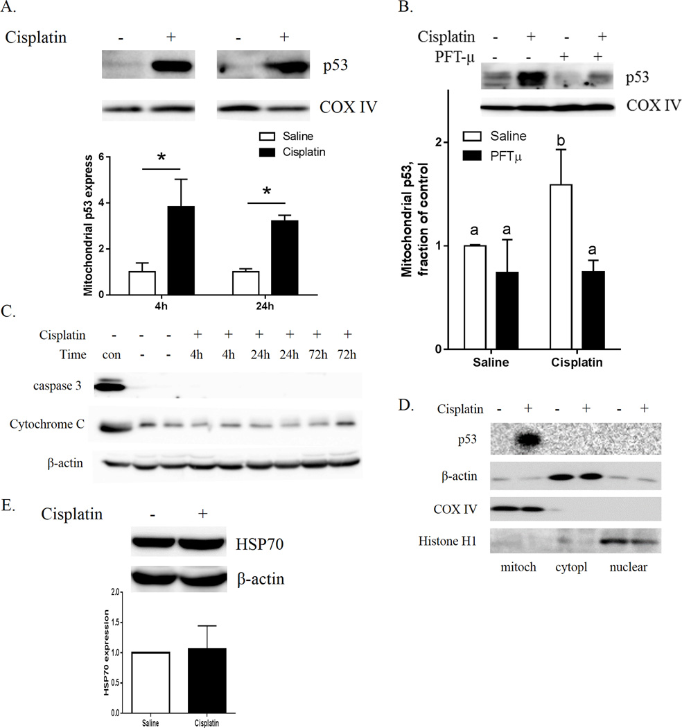
Figure 4. Acute effects of cisplatin on p53 translocation to the mitochondria and caspase-3 activation.
(A) Mitochondrial p53 was measured 4 h and 24 h after cisplatin injection and (B) 4 h after cisplatin and PFT-µ injections. COX-IV was used as a protein loading control. Relative expression of p53 was calculated 4 h and 24 h after cisplatin treatment. (C) The cytosolic fractions were analyzed for the apoptotic markers cytochrome-c and cleaved caspase-3 4 h, 24 h, and 72 h after a single dose of cisplatin. “Con” designates positive controls: brain mitochondria fraction for cytochrome-c (upper panel), and whole-cell lysate of H2O2-treated N2A cells for active caspase-3 (middle panel). β-actin was used as a protein loading control for cytosolic proteins. (D.) Cisplatin increases mitochondrial p53 without inducing p53 in cytosol or nucleus. Subcellular fractions collected at 4 h after cisplatin injection were analyzed for p53, Cox-IV (mitochondria), β-actin (cytosol and mitochondria) and Histone H1 (nuclear fraction). (E) Cytosolic HSP70. β-actin was used as a protein loading control. Results are expressed as means ± SEM; n = 6. *P < 0.05. Bars without a common superscript are significantly different (P < 0.05).

