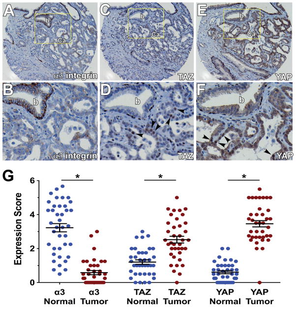
Figure 7. Loss of α3 integrin and increased nuclear TAZ and YAP in human prostate cancer specimens.
Immunohistochemical staining of α3 integrin (A&B), TAZ (C&D), and YAP (E&F) on adjacent sections of a prostate cancer tissue microarray. B, D, and F show enlarged views of the indicated fields in A, C, and E (b, benign gland; Ca, prostate carcinoma). Arrowheads indicate nuclear TAZ and YAP staining in prostate carcinoma. (G) Mean staining scores for α3 integrin, TAZ, and YAP on a 40 case microarray of human prostate cancer and matched normal tissues. Each case was represented by 4 cores of cancer and 4 cores of normal tissue. Each point represents the average score for each patient sample, across the 4 cores. The expression of α3 integrin was reduced, while nuclear expression of TAZ and YAP was increased in tumor versus normal glands *P<0.0001, paired t test. For scoring TAZ and YAP in normal glands, only luminal cells were considered.

