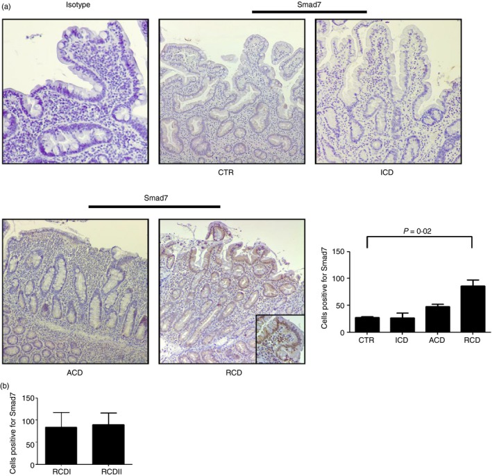
Figure 2.

Smad7 expression is increased in both epithelial and lamina propria compartments in refractory coeliac disease (RCD). (a) Representative photomicrographs (×100 original magnification) of Smad7‐stained paraffin‐embedded sections of biopsy samples taken from one control (CTR), one patient with active coeliac disease (ACD), one patient with inactive coeliac disease (ICD) and one patient with RCD. In RCD, Smad7‐positive cells are evident in both the epithelial and lamina propria compartments. Higher magnification photomicrograph (× 200) is shown in the insert. Staining with control IgG is also shown. Right panel shows the number of Smad7‐positive cells per high‐power field (hpf) in duodenal sections taken from three CTR, four patients with ACD, four patients with ICD and seven patients with RCD (four RCDI and three RCDII). Data indicate mean ± SE. (b) Number of Smad7‐positive cells in duodenal sections of four patients with RCDI and three patients with RCDII. Data indicate mean ± SE. [Colour figure can be viewed at wileyonlinelibrary.com]
