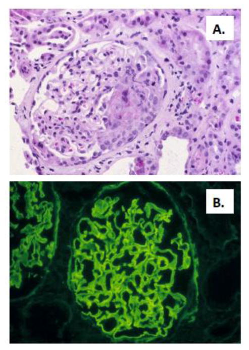
Figure 1.
Renal biopsy specimen from a patient with anti-GBM GN. A. Light microscopy showing focal and segmental necrotizing crescentic GN, with a segmental cellular crescent. B. Direct immunofluorescence showing intense linear staining for IgG along the glomerular capillary walls. Photomicrographs are courtesy of David N. Howell, MD PhD, Department of Pathology, Duke University Medical Center.

