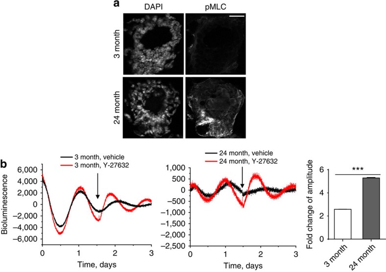
Figure 10. The mechano-sensing pathway influences the mammary clock in vivo.
(a) IF staining of pMLC expression in either 3-month or 24-month-old mammary gland tissue. N=3 animals, scale bar=50 μm. (b) Representative PER2::Luc bioluminescent traces of mammary tissue explants from 3-month or 24-month-old mice, treated with Y-27632 (100 μM, black arrow). Red, Y-27632 treatment; black, control treatment. Fold change of amplitude was quantified based on first peak following the treatment. Student's t-test, mean±s.e.m., ***P<0.001. N=3 animals.

