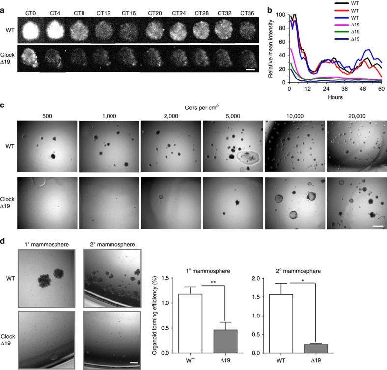
Figure 3. Mammary stem cells contain functional clocks that are required for their self-renewal capacity.
(a) Images of time lapse PER2::Luc bioluminescence in mammosphere structures isolated from WT or ClockΔ19 mutant female mice. Images represent sequential circadian time points after synchronisation with Dex. CT 0 (circadian time 0) denotes 24 h post Dex treatment. Note that owing to the weak signals from the ClockΔ19 mutant cells, the bioluminescence signals of the bottom panel were enhanced by 4 × to ease visualisation. N=3 animals. Scale bar=20 μm. (b) Quantification of mean intensity of PER2::Luc activity from individual wild-type or ClockΔ19 mammosphere structures. Three representative traces were plotted for each group. (c) Limiting Serial Dilution Analysis was performed using primary MECs isolated from wild-type or ClockΔ19 mutant mice. Numbers of single cells plated per cm2 is shown at the top. N=4 animals, scale bar=100 μm. (d) Primary mammosphere assay was performed at a cell density of 2,000 individual cells cm−2 from wild-type or ClockΔ19 mutant mice. Then a secondary mammosphere assay was performed by seeding 2,000 individual cells cm−2 from dissociated primary mammospheres. Student's t-test, data were shown as mean±s.e.m. *P< 0.05; **P< 0.01, n=4 animals. Scale bar=100 μm.

