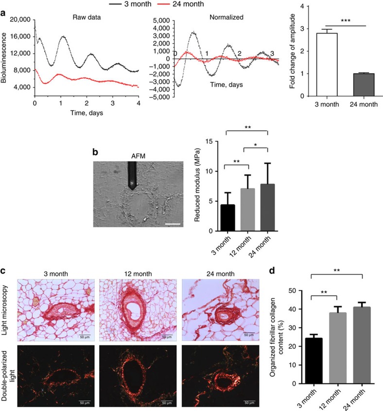
Figure 4. Aged mammary gland has a dampened clock and a stiffer mechano-microenvironment.
(a) Representative PER2::Luc traces from mammary tissues of either 3-month- (black) or 24-month-old (red) female mice. Right: fold change of amplitude was quantified (***, P<0.001, n=5 animals). (b) AFM measurements of periductal stiffness. Left: brightfield image of an unstained section under the AFM microscope with the periductal stroma clearly visible under the tip of the cantilever. Right, AFM quantification between 3-, 12- and 24-month-old mammary tissues. Data are shown as mean±s.e.m., **P<0.01, *P<0.05, n=3 animals, 1,200 replicates. Scale bar=50 μm. The reduced modulus is related to the Young's modulus but includes corrections for the compliance of the indenter when indenting biological substrates with soft tips. (c) Picrosirius red staining in brightfield condition (top panel) and under double-polarized light (bottom panel) of 3-, 12- and 24-month-old mammary gland sections. N=6 animals, scale bar=50 μm. (d) Percentage of organized fibrillar collagen in (c) was quantified. One-way ANOVA, mean±s.e.m., **P<0.01.

