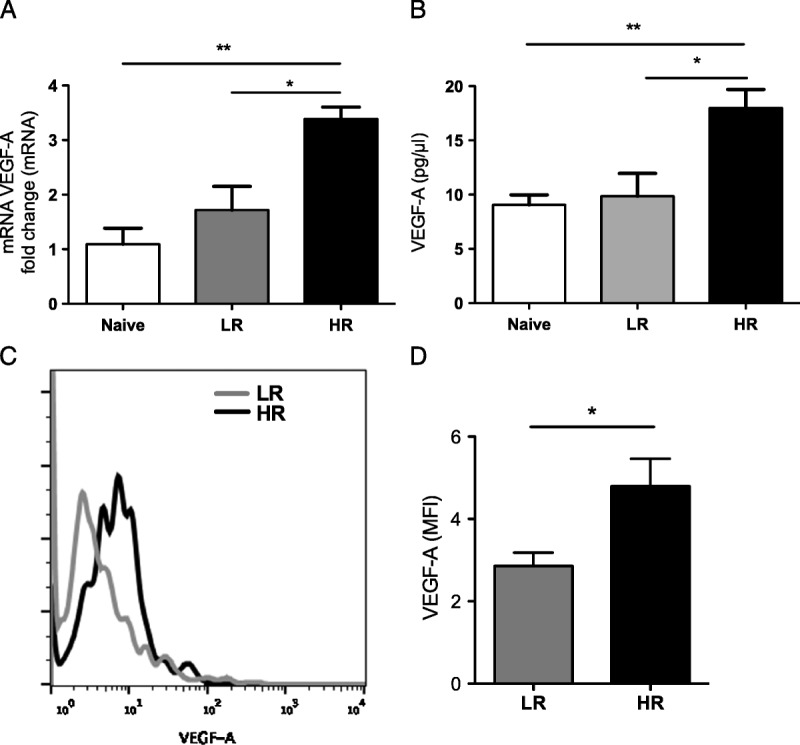
FIGURE 2.

Alloprimed T cells from high-risk mice release increased proangiogenic VEGF-A. A, T cells from naïve, LR, HR transplanted mice were stimulated with an anti-CD3 antibody for 12 hours. VEGF-A mRNA expression of T cells was assessed using real-time PCR and (B) VEGF-A protein concentration was detected in the culture supernatant using ELISA. Each experiment has been performed at least 3 times, with 10 mice per group per experiment. (C and D) Protein expression (median fluorescence intensity, MFI) of VEGF-A in corneal CD4 T cells isolated from HR and LR graft recipients 21 days posttransplantation was analyzed by flow cytometry (P = 0.04; n = 4/group).
