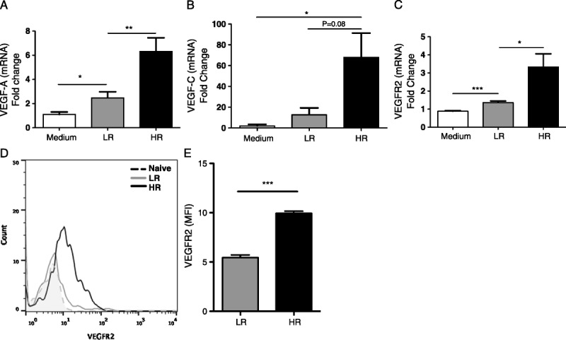
FIGURE 3.

VEGF-A, VEGF-C, and VEGF-R2 are increased in vascular endothelial cells. VECs were cultured alone (medium), or with T cells collected from LR or HR transplanted mice. mRNA expression of (A) VEGF-A, (B) VEGF-C, and (C) VEGF-Receptor 2 (VEGF-R2) was determined in VECs 24 hours after coculture. Each real-time analysis has been performed at least 3 times, with 10 mice per group per experiment. D and E, Flow cytometry analysis showing VEGF-R2 expression by CD31+ endothelial cells in the cornea of HR and LR transplanted mice 21 days postsurgery (P < 0.0001; n = 4/group).
