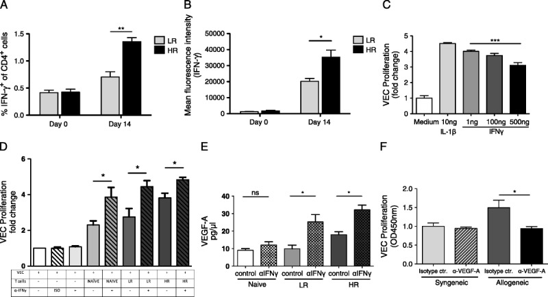
FIGURE 4.

Blocking IFNγ increases vascular endothelial cell proliferation. A and B, Before surgery (day 0) and 14 days after LR and HR corneal transplantation, draining lymph nodes were isolated and analyzed for (A) frequencies of IFNγ-producing T cells and (B) protein expression (MFI) of IFNγ in CD4 T cells. C, VECs were cultured in DMEM and stimulated with VEGF-A (20 ng/mL). Different concentrations of IFNγ (1, 100, 500 ng/mL) and IL-1β (10 ng/mL) were added for 24 hours. VEC proliferation was assessed using the BrdU incorporation assay. D, VECs were cultured in DMEM only, or with T cells from naïve, LR, or HR mice and treated with an anti-IFNγ or isotype control antibody as indicated. 24 hours later VEC proliferation was assessed using the BrdU incorporation assay. E, VEGF-A protein expression was assessed in the supernatant after culture using ELISA. F, Syngeneic and allogeneic T cells were cocultured with VECs and treated with anti-VEGF-A or isotype control for 24 hours. VEC proliferation was assessed using the BrdU incorporation assay. Each experiment has been performed at least 3 times, with 10 mice per group per experiment.
