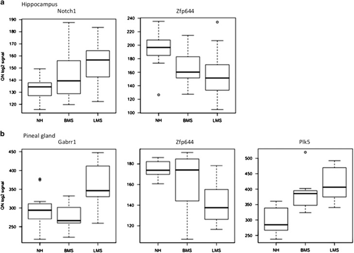
Figure 1.
Box plots illustrating relative expression levels (quantile normalized, log2-transformed signal intensities) of Notch1, Zfp644, Gabrr1 and Plk5 in hippocampus (a) and/or pineal gland (b) of rat offspring experiencing long (LMS), brief (BMS) or no (NH) maternal separation. The box plots indicate the median of the distribution (thick black line), 75th percentile (upper edge of box), 25th percentile (lower edge of box), 95th percentile (upper edge of vertical line), 5th percentile (lower edge of vertical line) and the outlier points (above and below vertical lines).

