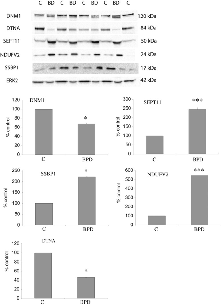
Figure 2.
Validation of differentially expressed proteins. Protein expression changes were determined in the 18 subpooled cases of the Stanley Medical Research Institute Array Collection using western blot analysis. Representative images and the means of three independent experiments are presented. Error bars indicate s.d. Western blots were prepared using lysates of subpools of anterior cingulate cortex samples from patients with bipolar disorder (BD or BPD) and control subjects (C). Immunoblots were incubated with antibodies that specifically recognize Dynamin (DNM1) at 120 kDa, Dystrobrevin (DTNA) at 84 kDa, Septin 11 (SEPT11) at 50 kDa, (NDUFV2) at 24 kDa, single-stranded DNA-binding protein (SSBP) at 17 kDa and ERK2, used as a loading control, at 42 kDa. The images show a typical blot and the corresponding graphs represent the signal intensity of the designated antibody measured by densitometry and corrected by the signal intensity of ERK2. The mean of three independent experiments is presented. Error bars indicate s.d. ERK2 showed no significant differences between disease and control (P=0.66). *P<0.05, ***P<0.001. In keeping with our liquid chromatography–mass spectrometry (LC-MS/MS) experiments, DNM1 and DTNA1 expression levels were reduced and SEPT11 and NDUF2 were found to be increased. For DNM1, bipolar disorder cases showed two bands, indicating potential post-translational modifications of the protein. However, we measured the band at 120 kDa for consistency. SSBP was found to be decreased in the MS data but increased by western blotting. This most probably is due to the antibody recognizing different/all isoforms of the protein.

