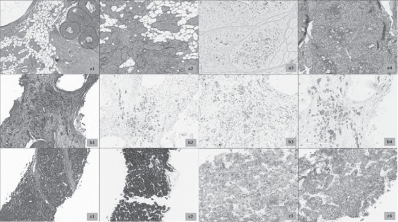
Fig. 1.
a Hematoxylin and eosin-stained (H&E) section of primary breast cancer, 1984 (1) signed-out as ‘malignant carcinoid’. Note the presence of an in situ component with same cytological details (2) and positivity of Grimelius silver stain (3). Morphology of relapse occurring in 1987 is strictly similar, with more pronounced pseudorosettes formation. b H&E section of bone metastasis (1). Cancer nests showed immunohistochemical positivity for estrogen receptors (2), very low proliferative rate with Ki 67 (< 5%, 3) and 2+ score of HER2/neu (ASCO/CAP guidelines 2013, 4). c Biopsy of pelvic mass shows invasive carcinoma with neuroendocrine morphology (1), with intense positivity to synaptophysin (2) and chromogranin-A (2). HER2/neu was confirmed as 2+ score (4). Note that granular positivity of chromogranin-A is very similar in distribution to the Grimelius positivity observed in 1984.

