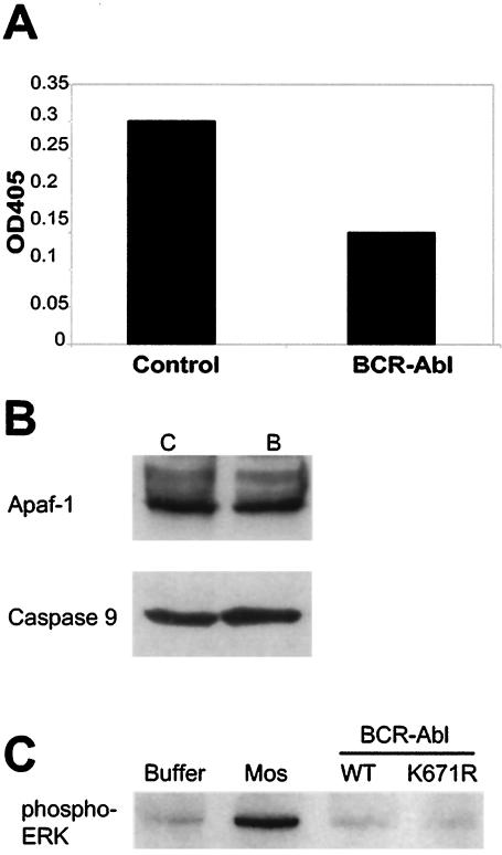
FIG. 4.
Bcr-Abl post-cytochrome c protection in cell lysates occurs by a posttranslational mechanism. (A) Control GFP-expressing cell lysates were preincubated with Abl immunoprecipitates from control GFP- or Bcr-Abl-expressing Rat-1 cells, after which caspase activity was assessed following the addition of 1 ng of soluble cytochrome c per μl. Caspase 3 activity was measured by assessing the cleavage of the colorimetric DEVD-pNA substrate. The results shown are representative of three independent experiments. (B) Control GFP (lane C)- and Bcr-Abl (lane B)-expressing Rat-1 lysates were immunoblotted for the Apaf-1 and caspase 9 proteins. (C) Cytosolic Xenopus egg extracts were pretreated with Mos, WT Bcr-Abl (WT), or kinase-dead Bcr-Abl (K671R). ERK activation was assessed by Western immunoblot analysis with an antibody directed against phospho-ERK. OD405, absorbance reading at 405 nm.

