Figure 1.
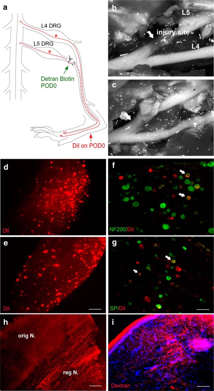
Anatomical evidence that the L5 spinal nerve regenerates in the SNL model. a, Schematic of the SNL model showing the site of L5 spinal nerve ligation and transection distal to L5 DRG and proximal to its merger with the L4 spinal nerve to form the sciatic nerve. The dotted line indicates the regenerated nerve segment. In separate experiments, the tracer dextran biotin (injected into the cut end of the spinal nerve) or DiI (injected into the paw) was injected just after the spinal nerve transection. b, c, In situ dissecting microscope images showing the regenerated segment of the L5 spinal nerve (arrows) just distal to the injury sites observed on day 20 (b) and day 70 (c). d, e, Whole-mount images of the L5 DRG showing DiI transported from the hindpaw (red) on day 20 (d; observed in only one of four rats) and day 70 (e; observed in six of six rats). f, g, DRG sections showing colocalization (yellow; arrows) of DiI (red) with labeling for NF200 (f; green; marker for myelinated cells; repeated in five rats) or Substance P (SP; g; green, marker for nociceptive cells; repeated in five rats). Sections were obtained 70 d after spinal nerve ligation and transection. DiI could still be observed at the paw injection site at this time. h, Longitudinal section of sciatic nerve distal to the original transection site, just below the knee, showing dextran biotin-positive fibers 70 d after spinal nerve transection (repeated in four rats). The regenerating, dextran biotin-positive nerve (“reg N.”) appeared somewhat segregated from the dextran biotin-negative original intact nerve (“orig N.) i, Cross-section of paw skin, showing dextran biotin-positive fibers 35 d after spinal nerve transection (repeated in five rats). Scale bars: d, e, 250 µm; f–i, 100 µm.

