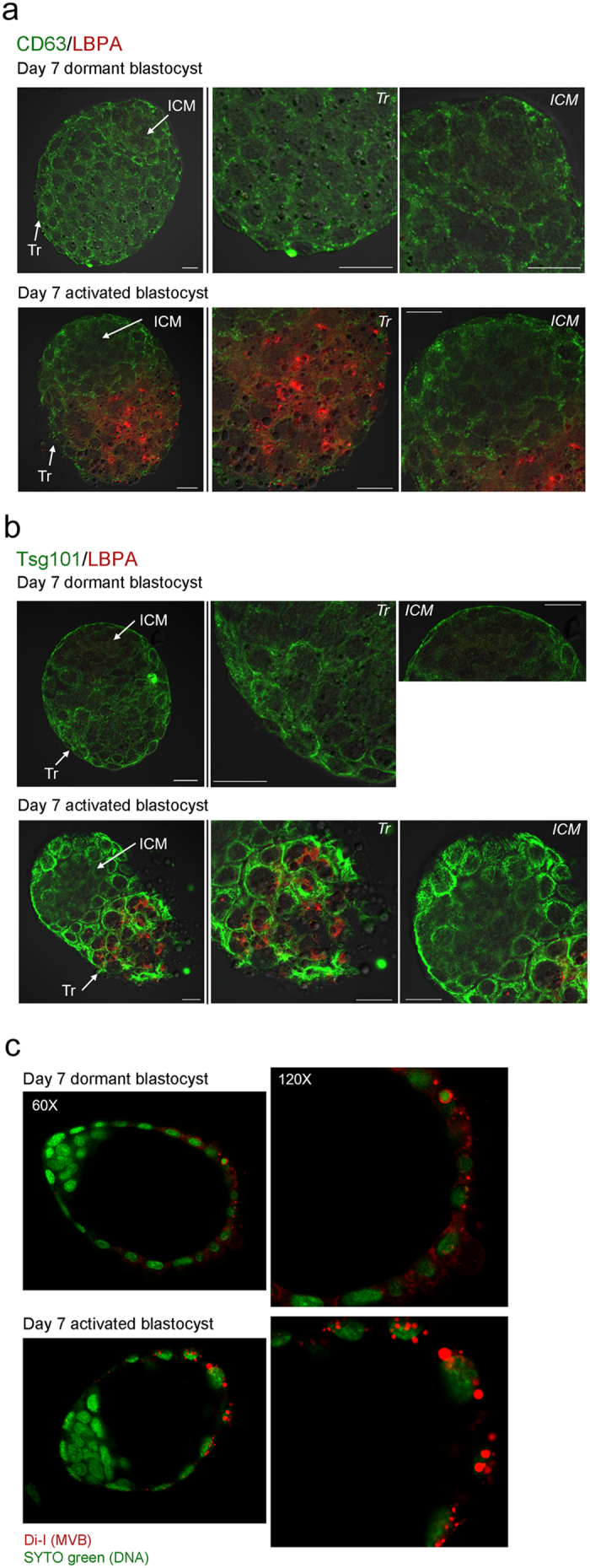
Figure 2. Numerous MVBs form in the trophectoderm of activated blastocysts.

(a) Immunofluorescence staining of CD63 and LBPA in dormant and activated blastocysts. Day 7 dormant and activated blastocysts were obtained and subjected to immunofluorescence staining. In both dormant and activated blastocysts, CD63 (green) exhibited a uniform puncta pattern throughout the trophectoderm (Tr). In contrast, LBPA (red) accumulation prominently increased in the mural trophectoderm of activated blastocysts where the blastocyst makes the initial contact with the endometrium during implantation. LBPA was not observed in the inner cell mass (ICM). Mouse or rabbit IgG was used as a mock control (data not shown). CD63 staining was repeated three times in 16 dormant (4 mice) and 20 activated blastocysts (5 mice) with similar results. Scale bar, 20 μm. (b) Immunofluorescence staining of Tsg101 and LBPA in day 7 dormant and activated blastocysts. Compared to the basal level of Tsg101 expression (green) in dormant blastocysts, Tsg101 accumulation dramatically increased in the trophectoderm of activated blastocysts. LBPA accumulation (red) in the mural trophectoderm of activated blastocysts was again confirmed. Mouse or rabbit IgG was used as a mock control (data not shown). LBPA staining was repeated three times in 17 dormant (4 mice) and 15 activated blastocysts (4 mice) with similar results. Tsg101 staining was repeated twice in 15 dormant (4 mice) and 12 activated blastocysts (3 mice) with similar results. Scale bar, 20 μm. (c) Confocal live imaging of Di-I stained embryos. Day 7 dormant and activated blastocysts were stained with dye (red) to stain internal vesicles of MVBs31. Nuclei were counterstained with SYTO 11 green fluorescence nucleic acid stain. Numerous Di-I-positive puncta are shown in the trophectoderm of activated blastocysts. The experiment was repeated three times with 28 dormant and 23 activated blastocysts.
