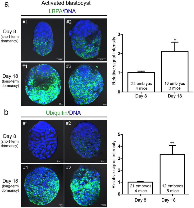
Figure 4. Accumulation of ubiquitin and LBPA increases in activated blastocysts after long-term delayed implantation.
Immunofluorescence staining was performed in short-term activated (activated on day 8) and long-term activated blastocysts (activated on day 18). (a) Immunofluorescence staining of LBPA was performed in indicated numbers of blastocysts for each group. Four independent experiments were performed and three sets showing similar results were included in statistical analysis (see Methods). Signal intensities were obtained by setting the whole blastocyst as the region of interest. The mean of values obtained for day 8 activated blastocysts in each experiment was normalized to 1. Two representative embryos (#1 and #2) are shown for each group. Nuclei were counterstained with TO-PRO-3 Iodide stain (blue). Scale bar, 20 μm. *p < 0.01. (b) Immunofluorescence staining of ubiquitin was performed in indicated numbers of blastocysts for each group. Four independent experiments were performed and were included in statistical analysis. Signal intensities were obtained by setting the whole blastocyst as the region of interest. The mean of values obtained for day 8 activated blastocysts in each experiment was normalized to 1. Both LBPA and ubiquitin signals were significantly higher in day 18 activated blastocysts. Two representative embryos (#1 and #2) are shown for each group. Nuclei were counterstained with TO-PRO-3 Iodide stain (blue). Scale bar, 20 μm. **p < 0.001.

