Figure 5.

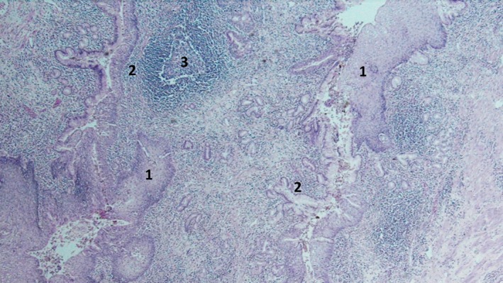
Cyst lined by mature squamous epithelium (1) continued by either columnar (2) or at places by cuboidal epithelium. Background shows diffuse inflammation with lymphoid follicles (3) surrounding adnexal‐type glands.

Cyst lined by mature squamous epithelium (1) continued by either columnar (2) or at places by cuboidal epithelium. Background shows diffuse inflammation with lymphoid follicles (3) surrounding adnexal‐type glands.