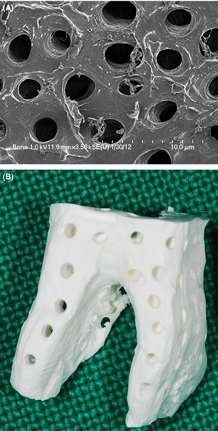
Figure 1.

Fabrication of the ABTB. (A) SEM of the processed ABTB surface. A clean surface and 1‐ to 5‐μm dentinal tubules provided space for protein exchange. (B) Lateral view of the ABTB. Macropores (200–300 μm) that penetrated from the surface to the pulp space provided the space for vascular invasion.
