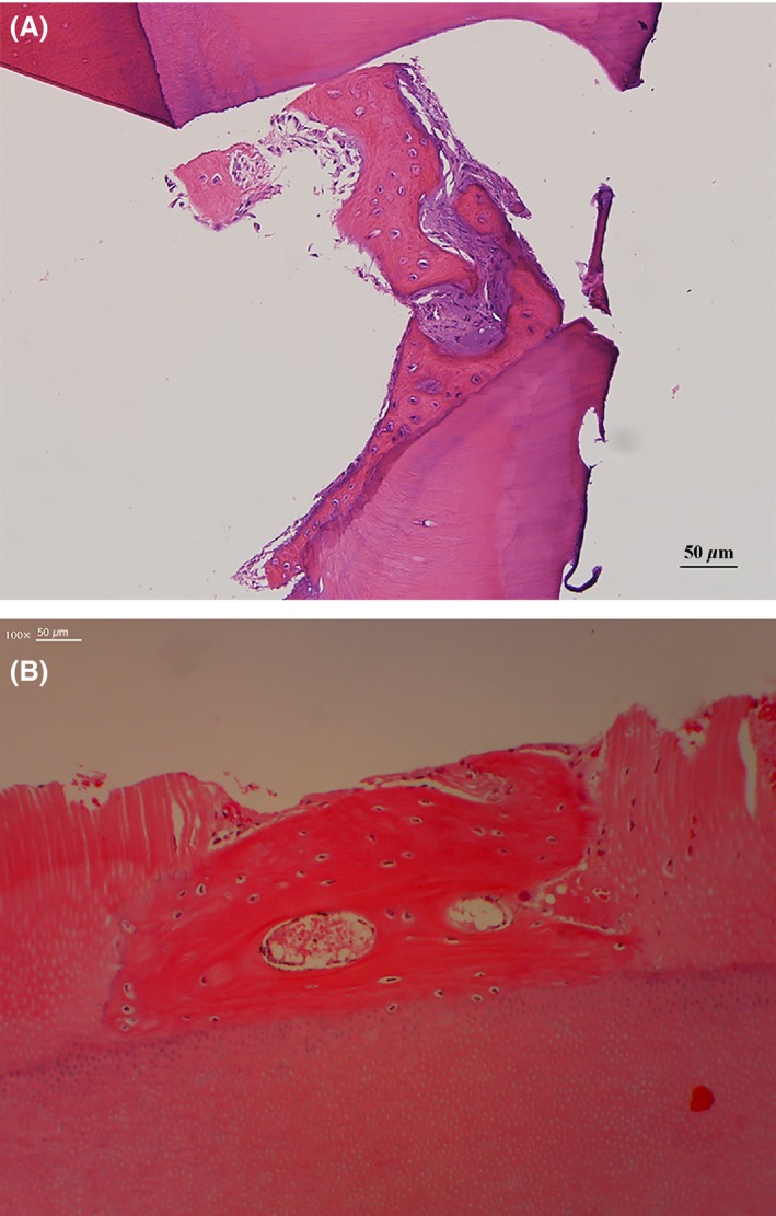
Figure 6.

Histological findings at the second surgery. (A) Histological specimen from maxillary case no. 6 in Table 1 at 8 months after the first surgery. A macropore of the ABTB was filled with newly formed osteoids with embedded active chondrocyte‐like cells that closely contacted the inner wall of the macropore. (B) Histological specimen from mandibular case no. 8 in Table 1 at 3 months after the first surgery. A newly formed osteoid, which had osteocytes and vessels, had been deposited on the ABTB surface. Cellular fusion without fibrous tissue invasion was observed on the border between the osteoid and the dentin matrix.
