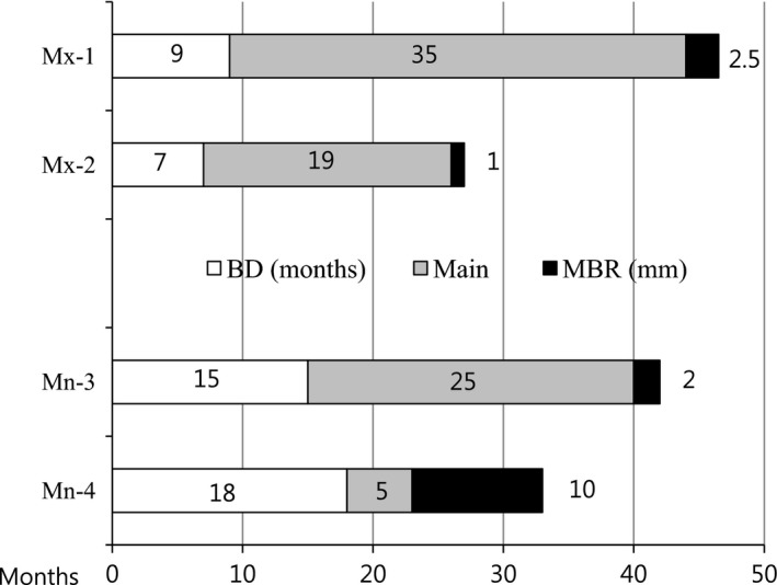
Figure 8.

MBR in the maxilla and the mandible. In the maxilla, MBRs of 2.5 and 1.0 mm were observed at 44 and 26 months after the first surgery, respectively. In the mandible, one patient exhibited 2.0 mm of MBR at #18 at 40 months after the first surgery, which featured a BD of 15 months. In particular, 10 mm of MBR indicated the removal of the 10‐mm implant at #18 due to the complete resorption of bone around the implant fixture (mandibular case no. 1). Mx, Maxilla; Mn, Mandible; Ma, Male; Fe, Female; BD, ABTB Disappearance; Main, maintenance; MBR, Marginal Bone Resorption.
