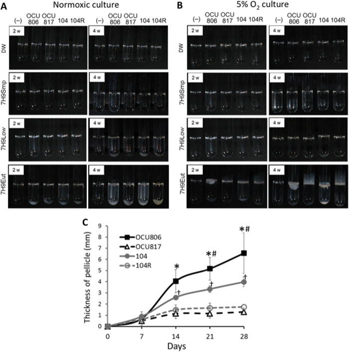
Figure 1. Observed difference in thickness and amount of pellicles between wild-type and rough mutant MAH strains.
(A,B) Pellicle formation in MAH. Bacteria were cultured in glass tubes for 4 weeks in normoxia (A) or 5% O2 condition (B) in distilled water (DW), simple 7H9 broth without supplementation of carbon and nitrogen sources (7H9Smp), 7H9Smp supplemented with 0.02% glycerol and 1% ADC enrichment (7H9Low), or 7H9Smp supplemented with 0.2% glycerol and 10% ADC enrichment (7H9Eut). (−) = no bacteria, 806 = MAH OCU806, 817 = MAH OCU817, 104 = MAH 104, and 104 R = MAH 104 R. (C) Time-course change of thickness of the pellicles in MAH cultured in 7H9Eut under 5% O2 condition. Data were expressed as means ± S.D from three independent experiments. *P < 0.01 versus the OCU817 group. #P < 0.05 versus the 104 group. †P < 0.05 versus the 104 R group.

