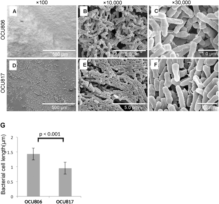
Figure 2. Ultrastructure of pellicles formed by wild-type and rough mutant MAH cells.
(A–F) Pellicles formed on air-liquid interface of 7H9Eut medium under 5% O2 condition at 3 weeks. High performance field is shown by enlarging area that contains bacterial clustering and extracellular matrix. (G) Comparison of the bacterial cell size (length of the major axis) between MAH OCU806 and MAH OCU817. Data were expressed as means ± S.D. (n = 50).

