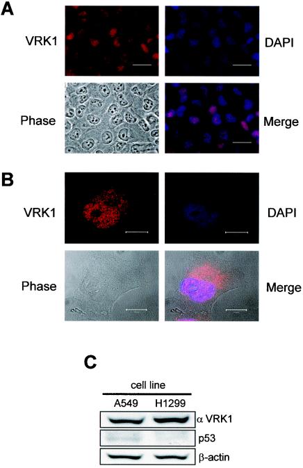
FIG. 1.
Localization of the endogenous human VRK1 protein in human A549 lung cancer cells determined by immunofluorescence (A) and confocal microscopy in interphase (B). The VRK1 protein was detected with the VC1 polyclonal antibody specific for the human protein labeled with Cy3. The nuclei were detected with DAPI staining, and the cells were identified by phase-contrast microscopy. The overlaps of the VRK1 and DAPI signals are shown in the merge panels. The bars represent 30 (A) or 10 (B) μm. (C) Immunoblot to detect endogenous levels of VRK1 and p53 in the two carcinoma cell lines used in the study using a polyclonal antibody specific for human VRK1 or anti-p53 monoclonal antibody.

