FIG. 8.
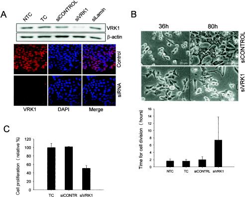
VRK1 suppression by specific siRNA provokes abnormalities in proliferation. (A) HCT116 cells were transfected with the specific siRNA for VRK1 (siVRK1), nontargeting siRNA (siCONTROL), or lamin-targeting siRNA (siLamin). Then the cells were processed for Western blotting or immunofluorescence assay as previously described with a specific antibody against endogenous VRK1 (red) or nuclear staining (DAPI). NTC, nontransfected cell control; TC, transfected cell control without siRNA. (B) After transfection as for panel A, cells were submitted to in vivo video microscopy. Representative images of cells 36 and 80 h after transfection are shown. The durations of cell division (plus standard deviations) were determined for 20 to 40 cells from several different experiments (below). (C) Quantification of cell proliferation and viability determined 60 h after transfection by colorimetric XTT-based assay.

