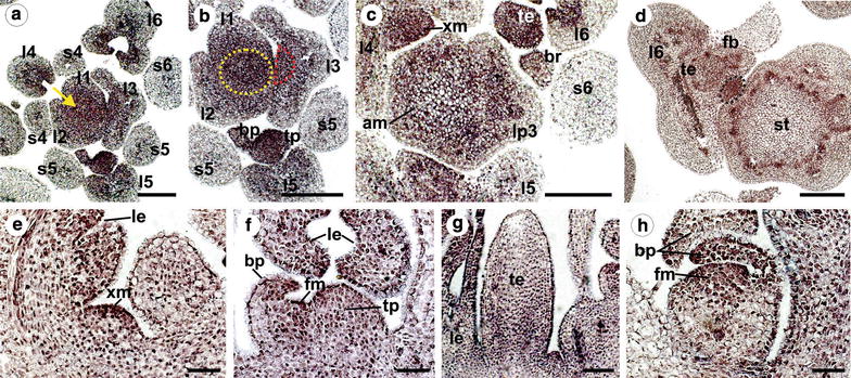
Fig. 6.

PeAP1 expression pattern by in situ hybridization of shoot apices of plants in the reproductive stage. a–d Cross sections of the P. edulis shoot apex from top to bottom. PeAP1 expression is detected in the adaxial side of the young leaves (l1–6), in the apical meristem (yellow arrow in a and yellow dashed circle in b), in the axillary meristem (dashed red semicircle in the third leaf primordium in b and “xm” in c), and in the tendril and bract primordia (tp and bp, respectively). PeAP1 transcripts were not detected in stipules (s). In d, PeAP1 is detected in the developing vegetative bud (black dashed circle), located between the tendril–flower complex (te and fb) and the stem (st). As sections distance from the apical meristem to the stem, PeAP1 expression in this region becomes fainter and it is not evident in the differentiated stem (st in d). e PeAP1 expression in axillary meristem (xm) in longitudinal section. le Leaf. f Longitudinal section of a tendril primordium and emerging flower meristem. PeAP1 transcripts are detected in the adaxial region of the bract primordium (bp), in the floral meristem (fm) and in the tendril primordium (tp). g Longitudinal section with PeAP1 transcripts detected in both young leaf (le) and tendril (te). h Expression of PeAP1 in a young flower bud, showing transcripts in the floral meristem (fm) and in bract primordia (bp). Bars: a, b, c, d = 100 µm; e, f, g, h = 50 µm
