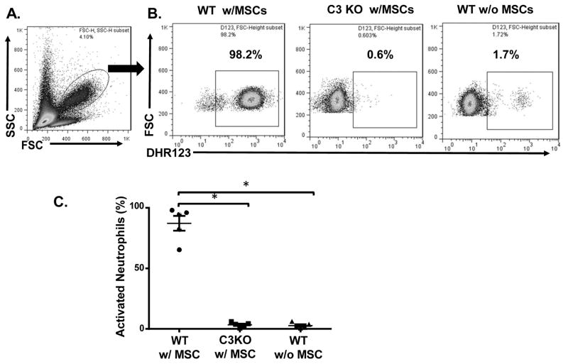
Figure 1. Neutrophils are activated by complement after MSC infusion.
MSCs were infused into WT or C3 KO mice (1×10⁶/mouse) by tail vein injection, then, after 40 min, peripheral blood was collected and activation of neutrophils in the blood assessed by staining the cells with DHR123, followed by flow cytometric analysis. WT mice without MSC infusion were included as controls. (A) Neutrophils in the peripheral blood. The oval gate indicates the cells analyzed in B and C. (B) Representative results showing activated neutrophils (DHR123+) in WT mice after MSC infusion (WT w/MSCs), C3 KO mice after MSC infusion (C3 KO w/MSCs), and control WT mice without MSC infusion (WT w/o MSCs). (C) Combined results for neutrophil activation assessment after MSC infusion. n=5 in each group and show the mean ± SD, One-way ANOVA and Tukey post-hoc test were used for data analysis, *p<0.05

