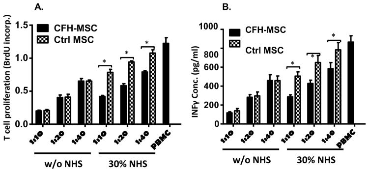
Figure 7. Painting CFH onto MSCs preserves their viability and function after contact with complement in vitro.
MSCs were painted with (CFH-MSCs) or without (Ctrl-MSCs) 10 μg of EDC-activated CFH, then were incubated for 30 min with 30% NHS in GVB++ (NHS) or GVB++ alone (w/o NHS). After washing and irradiation, 2×10⁴ of the MSCs were cultured in each well of a 96 well plate with different numbers of PBMCs in the presence of anti-CD3/CD28 beads and 30 U/ml of IL-2. After 48 h, 10 μM BrdU was added in each well, then, after 24 h, (A) the cells were harvested and proliferation of activated T cells by measuring levels of incorporated BrdU using a BrdU ELISA kit, and (B) supernatants were collected to measure IFNγ produced by the activated T cells. The data are results in triplicate representative of those in 4 independent experiments and are the mean ± SD, Two-way ANOVA tests were used in data analysis, *p<0.05

