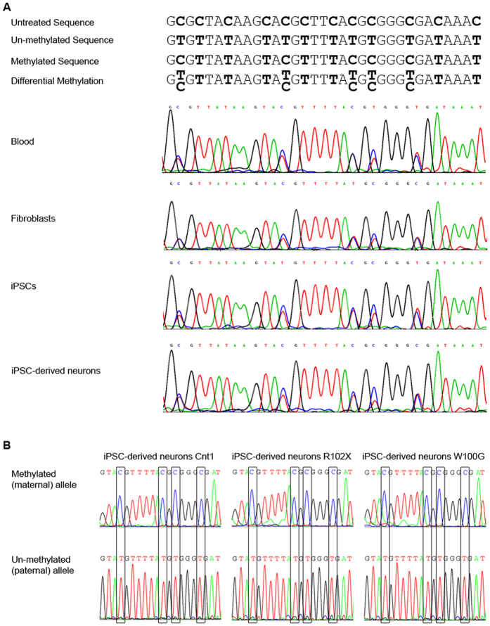
Figure 2. Detection of differentially methylated CpG dinucleotides in the promoter region of SGCE in iPSC-derived neurons.
The methylation pattern of the SGCE promoter was investigated by DNA sequencing with methylation-specific primers after bisulfite treatment. Upon bisulfite treatment, un-methylated cytosines are converted to uracil and therefore appear as thymines in the resulting sequence. (A) Possible sequencing outcomes and their interpretation are illustrated in the upper panel. In the lower panel, sequencing results of DNA extracted from blood, fibroblasts, iPSCs and iPSC-derived neurons of a healthy individual are shown. Differential methylation was detected in all tissues. (B) Methylation-specific sequencing of the SGCE promoter region in iPSC-neurons of one control and both M-D patients revealed the presence of fully methylated DNA alongside fully unmethylated DNA in the samples representing the maternal and paternal allele, respectively. Continuous methylation (i.e. no alternation of non-methylated and methylated CpG islands) was detected.

