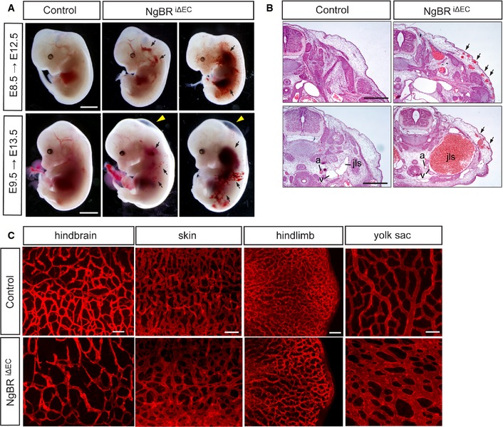
Figure 2. Hemorrhages and reduced capillary density in inducible NgBR endothelial‐specific knockout embryos.

- Whole‐mount view of NgBR i∆ EC mutant and control embryos at E12.5 and E13.5 after tamoxifen administration at E8.5 and E9.5, respectively. Hemorrhagic lesions (arrows) and subcutaneous edema (yellow arrowheads) were observed in NgBR i∆ EC mutants. Scale bar, 2 mm.
- Histological analysis of NgBR i∆ EC mutant and control embryos at E13.5 after tamoxifen administration at E9.5. Cross sections of the embryos were stained with hematoxylin and eosin. a, aorta; v, vein; jls, jugular lymph sac. Scale bar, 1 mm.
- Immunofluorescence staining with CD31 on various tissues in NgBR i∆ EC mutant and control embryos at E12.5 after tamoxifen administration at E8.5. Scale bar, 100 μm.
