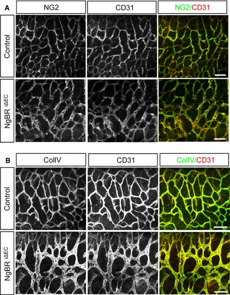
Figure EV2. Normal pericyte coverage and no vessel regression in the NgBR i∆ EC embryo.

- NG2 immunostaining with CD31 on the hindbrain of NgBR i∆ EC embryos at E12.5. There was no obvious difference in NG2‐positive pericyte distribution between control and NgBR i∆ EC.
- Immunodetection of collagen IV and CD31 in the hindbrain of NgBR i∆ EC embryos at E12.5. Vessel regression that is associated with the presence of empty basement membrane sleeves was not detected in NgBR i∆ EC.
Data information: Scale bar, 100 μm.
