-
A–D
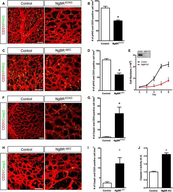
Immunofluorescence (IF) staining with CD31 (red) and pHH3 (green) on E9.5 NgBRECKO yolk sac and (B) quantification of pHH3‐positive cells. (C) IF staining with CD31 (red) and pHH3 (green) on E12.5 back skin of NgBR
i∆
EC and control embryos. (D) Quantification of (C).
-
E
Reduced proliferation of MLEC by NgBR deletion. MLEC isolated from NgBR
f/f animals were infected with Ad‐GFP (Control) or Ad‐Cre (NgBR KO). Cells were counted for 5 days after infection (n = 3). NgBR deletion in MLEC was detected by Western blotting with NgBR antibody.
-
F–I
IF staining for cleaved caspase‐3 on E9.5 NgBRECKO yolk sac (F) and E12.5 back skin of NgBR
i∆
EC embryos (H) and respective quantification of Casp3‐positive cells (G, I).
-
J
Caspase‐3 activity assay on control and NgBR KO MLEC.
Data information: Scale bar, 100 μm. *
P < 0.05.
P‐values were calculated by unpaired Student's
t‐test. Data are mean ± SEM,
n = 7–12 per group.

