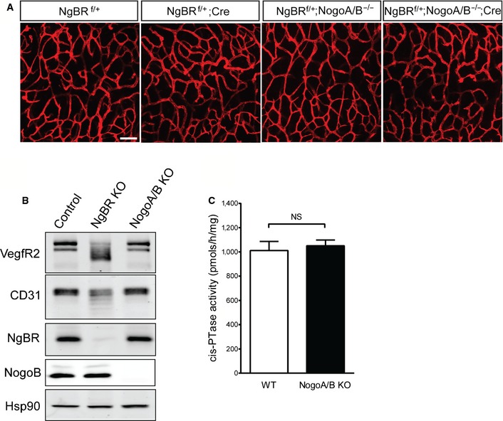
Figure EV4. Nogo‐B is not required for NgBR functions in endothelial cells during embryogenesis.

- Staining with anti‐CD31 on hindbrain of E12.5 embryos after tamoxifen injection at E8.5. Scale bar, 100 μm.
- Western blot analysis for protein glycosylation in MLEC. Nogo‐A/B KO MLEC were isolated from Nogo‐A/B KO animals. Expression of VEGFR2 and CD31 was detected. Knockout cells were confirmed by detection with anti‐NgBR and anti‐Nogo‐B antibodies. Hsp90 was used as a loading control.
- Microsomal cisPTase activity assay for Nogo‐A/B KO MEF. No significant difference was detected between control and NogoA/B KO cells. cisPTase activity assay was performed as described in 4. NS, not significant (P > 0.05). P‐value was calculated by unpaired Student's t‐test. Data are mean ± SEM, n = 3 per group.
