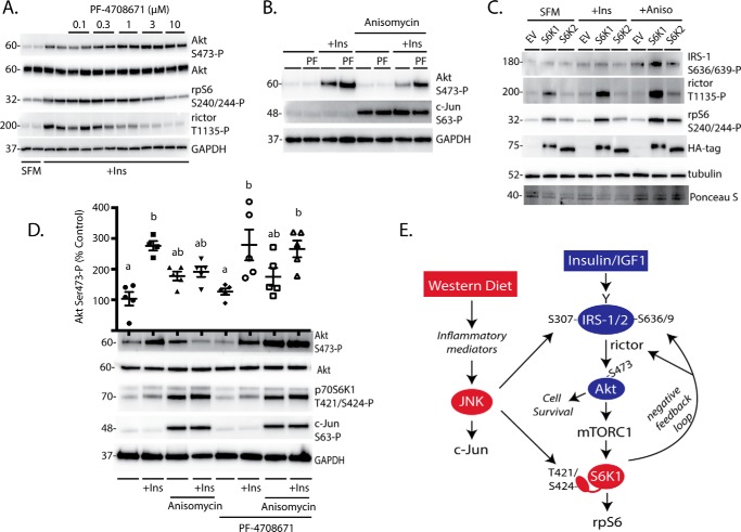
FIGURE 7.
Inhibition of p70S6K1 maintains insulin action in retinas upon JNK activation. A, HEK293E cells were serum-deprived for 2 h and treated with 0.1–10 μm PF-47086 for the last 30 min to inhibit p70S6K1. Cells were then treated with insulin (100 nm) for 30 min as indicated. B, cells were serum-deprived and treated with PF-47086 (10 μm) to inhibit p70S6K1. Cells were then treated with anisomycin and/or insulin as indicated for 30 min. C, S6K1/2 knock-out MEF were transfected with HA-tagged S6K1, HA-tagged S6K2, or an EV control followed by serum deprivation (SFM) and treatment with insulin and anisomycin as described above. Phosphorylation of rpS6 on Ser-240/244, rictor on Thr-1135, and eIF4B on Ser-422 as well as expression of Akt, IRS-1, GAPDH, and HA-tagged S6K1/2 were assessed by Western blotting. Blots shown in A–C are representative of three independent experiments; within each experiment three independent samples were analyzed. D, explant tissue culture was used to study insulin action in retina without the influence of the blood-retinal barrier. Mouse retinas were removed and incubated in tissue culture medium in either the presence or absence of PF-4708671 (10 μm) for 15 min. Retinas were treated with anisomycin (20 μm) and/or insulin (100 nm) as indicated for 30 min. Phosphorylation of Akt on Ser-473, p70S6K1 on Thr-421/Ser-424, and c-Jun on Ser-63 were evaluated by Western blotting. Values are the means ± S.E. Statistical significance is denoted by the presence of different letters above the scatter plots on the graphs. Scatter plots with different letters are statistically different; p < 0.05. Results in D are representative of two experiments; within each experiment, two or three independent samples were analyzed. Protein molecular mass in kDa is indicated at the left of the blots. E, working model for the mechanism whereby a Western diet attenuates insulin action in the retina.

